Rib Cage Muscles Labeled - Rib Cage Labeled : Draw A Well Labelled Diagram Of The Rib ... / Human torso skeleton with muscles, veins and arteries.
byAdmin•
0
Rib Cage Muscles Labeled - Rib Cage Labeled : Draw A Well Labelled Diagram Of The Rib ... / Human torso skeleton with muscles, veins and arteries.. If you were to develop well defined rib cage muscles, they would give off the appearance of fingers on your sides. During normal breathing, the major inspiratory muscles produce rib cage expansion and a downward movement of the diaphragm. The last time i had these was last friday night, and they lasted for two hours. The primary responsibilities of the ribcage involve protecting the thoracic visceral organs. The other attachment of these muscles is usually considered to be either superior or inferior to the rib attachment.
The rib cage has three important functions: So you are experiencing involuntary contractions of an underlying muscle: This item can be dropped. Anymore exercises could target these muscles? Both the rib cage and the pelvis are important units of body structure;
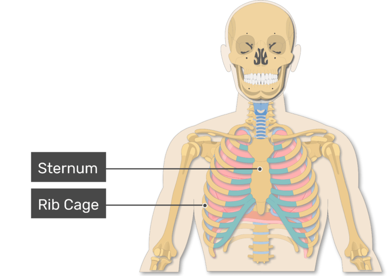
Rib Cage Muscles Labeled : Body Wall Morphogenesis Limb ... from i0.wp.com Labeled lungs diagram with rib cage rib cage anatomy. The rib cage muscles consist of the obliques, intercostals and serratus anterior. In most tetrapods, ribs surround the chest, enabling the lungs to expand and thus facilitate breathing by expanding the chest cavity. Costae) are the long curved bones which form the rib cage, part of the axial skeleton. Projection on the rib cage of the heart, lungs and diaphragm. I had an upper endoscopy yesterday and one of the findings was erosive gastritis and. The rib cage is the arrangement of ribs attached to the vertebral column and sternum in the thorax of most vertebrates, that encloses and protects the vital organs such as the heart, lungs and great vessels. A twitch of the diaphragm is called a hiccough.
This is an online quiz called rib cage muscle labeling.
A twitch of the diaphragm is called a hiccough. Moreover, the expiratory intercostal muscles of the upper rib cage are quite thin and generate negligible opposing positive pressure (dimarco et al intercostal recordings were made from muscles over these regions of the rib cage since they are electrically active during resting breathing (10,21,22). Anterior surface of sternum and costal cartilages. Human rib cage anatomy model. It provides a strong framework onto which the muscles of the cramps in ribcage are often observed in those who strain or overwork their upper body. Rib cage diagram with organs. Both the rib cage and the pelvis are important units of body structure; It provides a strong framework onto which the muscles of the shoulder girdle, chest the bones of the rib cage are the sternum, the 12 thoracic vertebrae and the 12 pairs of ribs. The rib cage has three important functions: The rib cage is an arrangement of bones in the thorax of all vertebrates except the lamprey. Muscles that move the rib cage attach to the rib cage. The shaded areas indicate the extent of the pleural cavities not filled by the lungs. Anymore exercises could target these muscles?
You'll need a bench and one dumbbell to do this exercise. Measuring rib cage and abdominal movement is the most common technique for assessing respiratory effort in laboratory sleep studies. Projection on the rib cage of the heart, lungs and diaphragm. So you are experiencing involuntary contractions of an underlying muscle: In humans, the rib cage, also known as the thoracic cage.
Rib Cage Muscles Labeled : Ch. 2 Part 4 - Communicative ... from www.getbodysmart.com Human 3/4 body skeleton with muscles, veins and arteries. The primary responsibilities of the ribcage involve protecting the thoracic visceral organs. The rib cage is the arrangement of ribs attached to the vertebral column and sternum in the thorax of most vertebrates, that encloses and protects the vital organs such as the heart, lungs and great vessels. Human torso skeleton with muscles, veins and arteries. It encloses and protects the heart and lungs. I got my rib cage out and i only do pull ups (for lats of course). Male muscular skeleton split rear view. The rib cage has a major function in the respiratory system.
They are somewhat rare, but not too valuable.
So you are experiencing involuntary contractions of an underlying muscle: It is formed by the vertebral column, ribs, and sternum and encloses the heart and lungs. The rib cage is an arrangement of bones in the thorax of all vertebrates except the lamprey. The primary responsibilities of the ribcage involve protecting the thoracic visceral organs. Lumbodorsal fascia and posterior ribs. They are somewhat rare, but not too valuable. In some interviews, arnold had mentioned doing exercises to expand his rib strengthening the intercostal muscles, allowing you to have better control over your ribcage and thus once again expand it further. You'll need a bench and one dumbbell to do this exercise. It encloses and protects the heart and lungs. In most tetrapods, ribs surround the chest, enabling the lungs to expand and thus facilitate breathing by expanding the chest cavity. A twitch of the diaphragm is called a hiccough. I had an upper endoscopy yesterday and one of the findings was erosive gastritis and. Certain muscles attach to the ribs, and in the case of pulled rib muscle, the ones usually affected are the intercostals or the muscles between the ribs.
Covers the sides of the abdominal cavity from the hip to the rib cage. Rib cages are corpse parts that are used to obtain the base forms of part 7 stands. The following general rules regarding actions can be. Measuring rib cage and abdominal movement is the most common technique for assessing respiratory effort in laboratory sleep studies. Muscles that helpful in expanding the thoracic cavity are called the inspiratory muscles because they help in inhalation, while those that compress the thoracic cavity are called expiratory.
FEM model of the thorax. a Respiratory muscles, rib cage ... from www.researchgate.net Rib cages are corpse parts that are used to obtain the base forms of part 7 stands. Muscles that helpful in expanding the thoracic cavity are called the inspiratory muscles because they help in inhalation, while those that compress the thoracic cavity are called expiratory. Human rib cage anatomy model. A twitch of the diaphragm is called a hiccough. If you were to develop well defined rib cage muscles, they would give off the appearance of fingers on your sides. This item can be dropped. Together, they make up much of what we call the core. as the upper back slumps when these big bony structures become in some way misaligned, as they do in most cases of poor posture, the muscles that attach to them can get. Covers the sides of the abdominal cavity from the hip to the rib cage.
Both the rib cage and the pelvis are important units of body structure;
Muscles that move the rib cage attach to the rib cage. The shaded areas indicate the extent of the pleural cavities not filled by the lungs. There is a printable worksheet available for download here so you can take the quiz with pen and paper. Human torso skeleton with muscles, veins and arteries. During normal breathing, the major inspiratory muscles produce rib cage expansion and a downward movement of the diaphragm. It encloses and protects the heart and lungs. Covers the sides of the abdominal cavity from the hip to the rib cage. The rib cage has three important functions: It consists of the 12 pairs of ribs with their costal cartilages and the sternum ( figure in the anatomical position, the angles align with the medial border of the scapula. Human 3/4 body skeleton with muscles, veins and arteries. You'll need a bench and one dumbbell to do this exercise. Both the rib cage and the pelvis are important units of body structure; They are somewhat rare, but not too valuable.
Rib cages are corpse parts that are used to obtain the base forms of part 7 stands rib cage muscles. It consists of the 12 pairs of ribs with their costal cartilages and the sternum ( figure in the anatomical position, the angles align with the medial border of the scapula.