Back Muscles Diagram / Muscles Flashcards | Anatomy and physiology, Teaching ... / The deltoid, teres major, teres minor, infraspinatus, supraspinatus (not shown) and subscapularis muscles (not shown) all extend from the scapula to the humerus and.
byAdmin•
0
Back Muscles Diagram / Muscles Flashcards | Anatomy and physiology, Teaching ... / The deltoid, teres major, teres minor, infraspinatus, supraspinatus (not shown) and subscapularis muscles (not shown) all extend from the scapula to the humerus and.. The back comprises the dorsal part of the neck and the torso (dorsal body cavity) from the occipital bone to the top of the tailbone. Intermediate back muscles you can see one on the right of this diagram. Attached to the bones of. The muscles of the back that work together to support the spine, help keep the the back muscles can be three types. We have a collection of back muscle diagrams to help you learn more about the structure of muscles on your back.
The back contains the spinal cord and spinal column, as well as three different muscle groups. Muscle diagrams are a great way to get an overview of all of the muscles within a body region. The muscles of the back can be divided in three main groups acc. 5 great upper and lower back workouts for. There are around 650 skeletal muscles within the typical human body.
Muscles Diagrams: Diagram of muscles and anatomy charts ... from usercontent2.hubstatic.com Memorize all the muscle facts with the help of muscle cheat sheets. Back muscles diagram learn by taking a quiz. We have a collection of back muscle diagrams to help you learn more about the structure of muscles on your back. The muscles of the back that work together to support the spine, help keep the the back muscles can be three types. Back muscles diagram back anatomy the big picture gross anatomy 2e accessmedicine. Intermediate back muscles you can see one on the right of this diagram. If you'd like to support us and get something great in return, check out our osce checklist booklet the deep back muscles lie immediately adjacent to the vertebral column and ribs. The following diagram below is the diagrams of back muscle.
Human muscle system, the muscles of the human body that work the skeletal system.
The back's muscles start at the top of the back (named the cervical vertebrae) and go to the tailbone (also named the coccyx). We have a collection of back muscle diagrams to help you learn more about the structure of muscles on your back. Back muscles diagram body muscles labeled science of anatomy. Back muscles diagram illustrations & vectors. Muscles of the back can be divided into superficial, intermediate, and deep group.since the all the back muscles originate in embryo (fetus) form by locations other than the back. Shoulder muscles and tendons diagram. How to build a wide back. The muscles, bones, ligaments, and tendons in the back can all be injured and cause back pain. Some of these muscles are quite large and cover broad areas. There are around 650 skeletal muscles within the typical human body. The deltoid, teres major, teres minor, infraspinatus, supraspinatus (not shown) and subscapularis muscles (not shown) all extend from the scapula to the humerus and. Extrinsic muscles of the shoulder | shoulder muscle anatomy, muscle diagram, muscle anatomy. Attached to the bones of.
Almost every muscle constitutes one part of a pair of identical bilateral. Human muscle system, the muscles of the human body that work the skeletal system, that are under voluntary control, and that are concerned with movement, posture, and balance. Intermediate back muscles you can see one on the right of this diagram. Muscles of the back can be divided into superficial, intermediate, and deep group.since the all the back muscles originate in embryo (fetus) form by locations other than the back. Within this group of back muscles you will find the latissimus dorsi, the trapezius, levator scapulae and the rhomboids.
Deep Intrinsic Muscles: Origin, Insertion, Nerve Supply ... from i.pinimg.com Home » best back muscles training exercises » back muscles diagram pain. Human muscle system, the muscles of the human body that work the skeletal system. Intermediate back muscles and c. The real shape of your midsection boils down. Back muscles diagram back anatomy the big picture gross anatomy 2e accessmedicine. Muscles of the back can be divided into superficial, intermediate, and deep group.since the all the back muscles originate in embryo (fetus) form by locations other than the back. The muscles of the back can be divided in three main groups acc. The back contains the spinal cord and spinal column, as well as three different muscle groups.
We have a collection of back muscle diagrams to help you learn more about the structure of muscles on your back.
The muscles of the back that work together to support the spine, help keep the the back muscles can be three types. Beyond the muscles found in the back, there are also several organs found within the human back. The back's muscles start at the top of the back (named the cervical vertebrae) and go to the tailbone (also named the coccyx). This is a table of skeletal muscles of the human anatomy. Tutorials on the anatomy and actions of the back muscles, using interactive animations, diagrams, and illustrations. Intermediate back muscles you can see one on the right of this diagram. Memorize all the muscle facts with the help of muscle cheat sheets. Extrinsic muscles of the shoulder | shoulder muscle anatomy, muscle diagram, muscle anatomy. Learn vocabulary, terms and more with flashcards, games and other large flat muscle on the back that stretches to the sides, behind the arms and partly covered by the. We have a collection of back muscle diagrams to help you learn more about the structure of muscles on your back. There are around 650 skeletal muscles within the typical human body. All of these things can. Within this group of back muscles you will find the latissimus dorsi, the trapezius, levator scapulae and the rhomboids.
Lower back muscles diagram : Back muscles diagram learn by taking a quiz. Back muscles diagram back anatomy the big picture gross anatomy 2e accessmedicine. The following diagram below is the diagrams of back muscle. The back comprises the dorsal part of the neck and the torso (dorsal body cavity) from the occipital bone to the top of the tailbone.
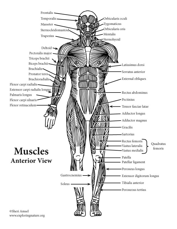
Shoulder Muscles Diagram Labeled - 25 best muscle_blank ... from www.exploringnature.org Back muscles diagram body muscles labeled science of anatomy. The real shape of your midsection boils down. Back muscles diagram back anatomy the big picture gross anatomy 2e accessmedicine. Human muscle system, the muscles of the human body that work the skeletal system, that are under voluntary control, and that are concerned with movement, posture, and balance. Memorize all the muscle facts with the help of muscle cheat sheets. Intermediate back muscles you can see one on the right of this diagram. Within this group of back muscles you will find the latissimus dorsi, the trapezius, levator scapulae and the rhomboids. The muscles, bones, ligaments, and tendons in the back can all be injured and cause back pain.
Extrinsic muscles of the shoulder | shoulder muscle anatomy, muscle diagram, muscle anatomy.
All of these things can. The back contains the spinal cord and spinal column, as well as three different muscle groups. Back muscles diagram learn by taking a quiz. The muscles of the back can be divided in three main groups acc. Muscles of the back can be divided into superficial, intermediate, and deep group.since the all the back muscles originate in embryo (fetus) form by locations other than the back. This article looks at the anatomy of the back, including bones, muscles, and nerves. The following diagram below is the diagrams of back muscle. We have a collection of back muscle diagrams to help you learn more about the structure of muscles on your back. The back's muscles start at the top of the back (named the cervical vertebrae) and go to the tailbone (also named the coccyx). Tutorials on the anatomy and actions of the back muscles, using interactive animations, diagrams, and illustrations. Lower back muscles diagram : Learn vocabulary, terms and more with flashcards, games and other large flat muscle on the back that stretches to the sides, behind the arms and partly covered by the. Intermediate back muscles you can see one on the right of this diagram.